According to the AHA – 1.5 million in the US suffer from Aortic Stenosis. Aortic valve stenosis (AS) is a complex disease caused by a narrowing or hardening of the aortic valve due to calcium buildup on the heart valve flaps.
Transcatheter aortic valve implantation (TAVI) has become an established treatment for severe aortic stenosis and is being performed for growing numbers of patients. It is estimated that > 250,000 TAVI procedures are performed globally each year.
Currently 25% of patients are unable to undergo TAVI procedure due to higher associated risks and complications from specific anatomical quirks.
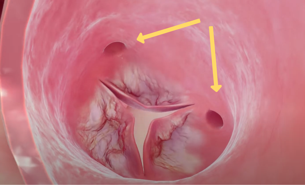
The orifice of the two coronaries (the arteries that supply blood to the heart muscle), is located behind the leaflets of the Aortic valve. There is an immediate risk of obstruction of the blood flow into the coronaries if the cusps of the replaced valve are pushed by the new implant.
This obstruction will immediately compromise the heart function placing the life of these patients at an instant and clear danger.
Top View: calcified leaflets and coronary orifice
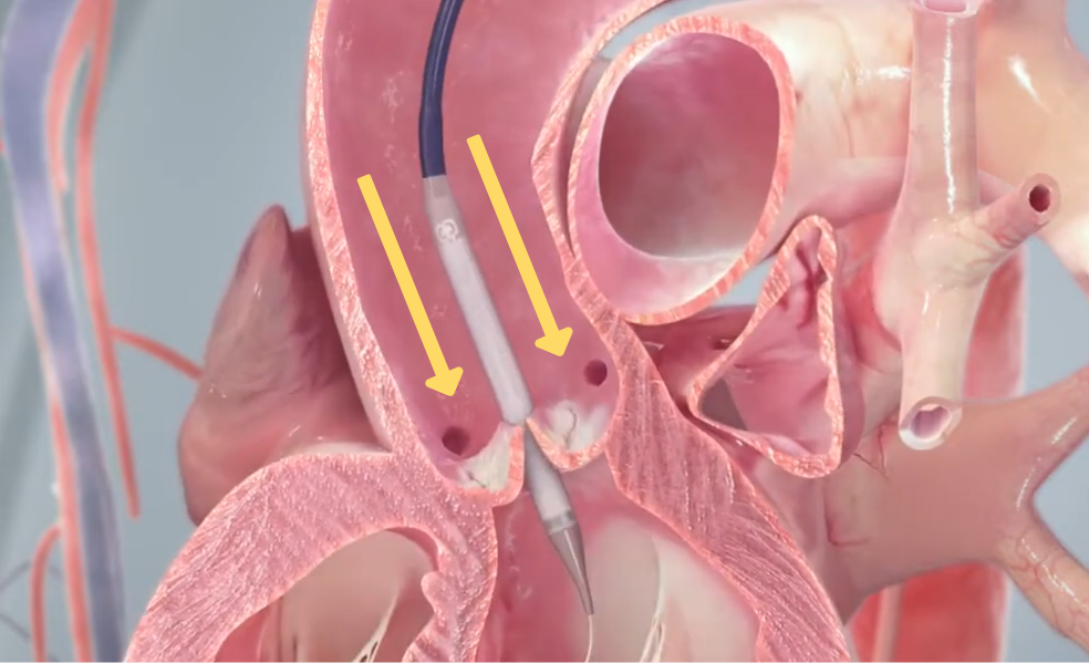
Side view: calcified leaflets
and coronary orifice, Tavi catheter
is in position
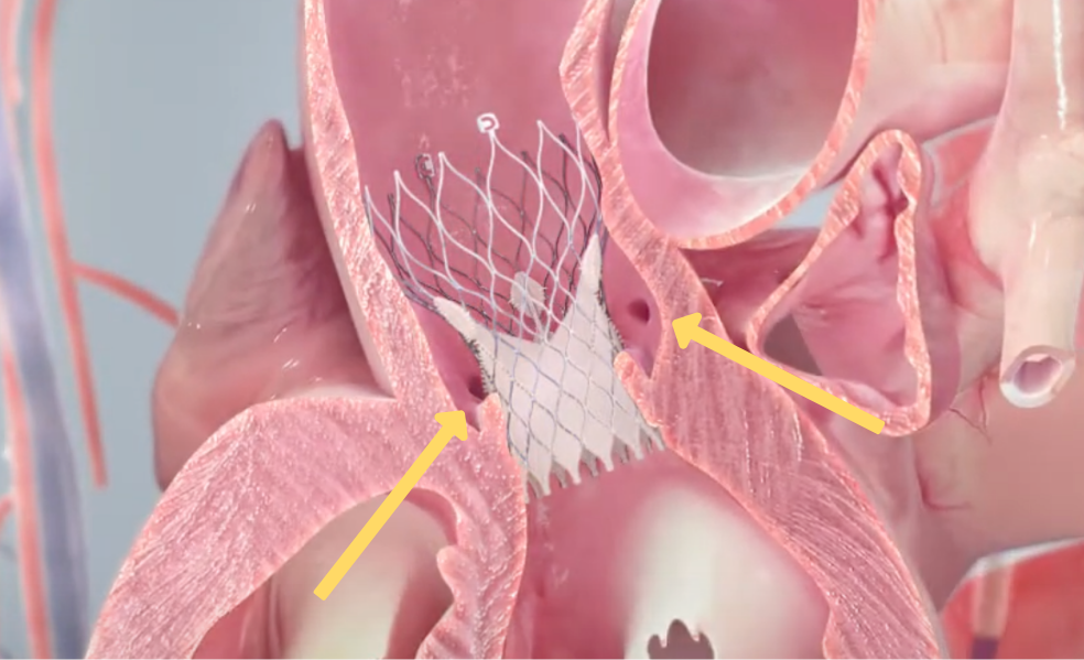
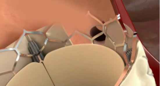
Side view: prosthetic valve is deployed.
Note the leaflets adjacent to the
orifice of the coronaries
The solution
HVT Medical’s Splitter device performs cusp splitting before a TAVI procedure, to help reduce risk of coronary occlusion.
The device creates an intentional laceration or excision of valve’s cusp tissue by applying an advanced catheter equipped with cutting head that mimics an alligator jaw.
The catheter rides a standard guide wire for TAVI operations so the splitter will be integrated completely into the workflow of the valve implementation.
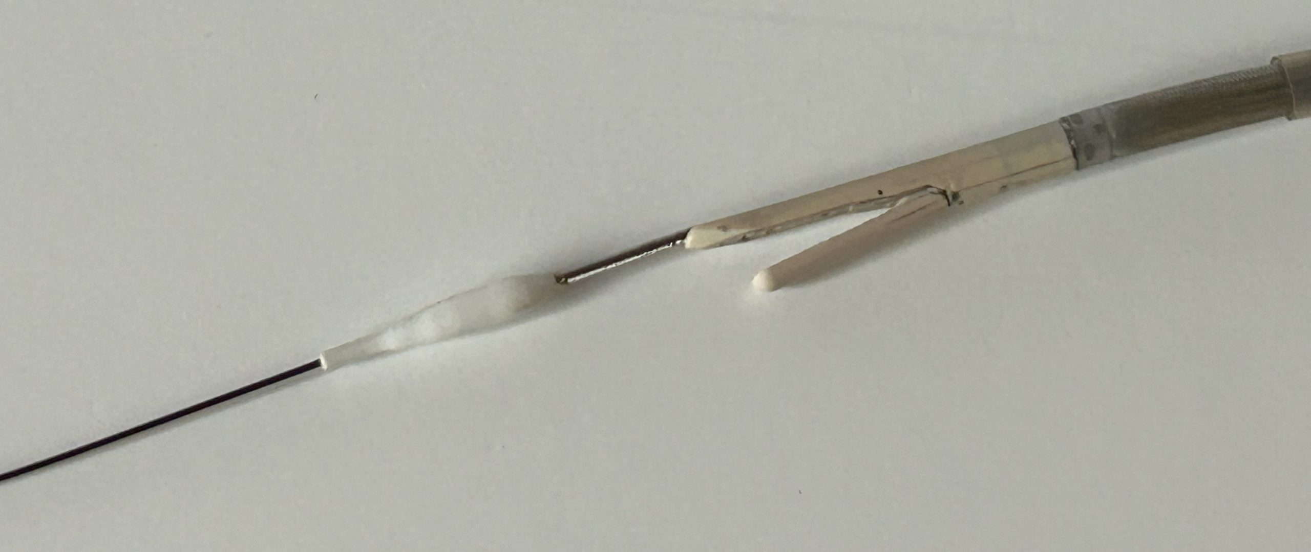
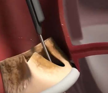
The a-traumatic nose cone is riding on a standard TAVI Guide wire. The 2 jaws of the cutting head are designed to grasp the selected leaflet and once closed, hold firmly the detached segment and remove it out of the body
The Nose Cone and the Cutting Head
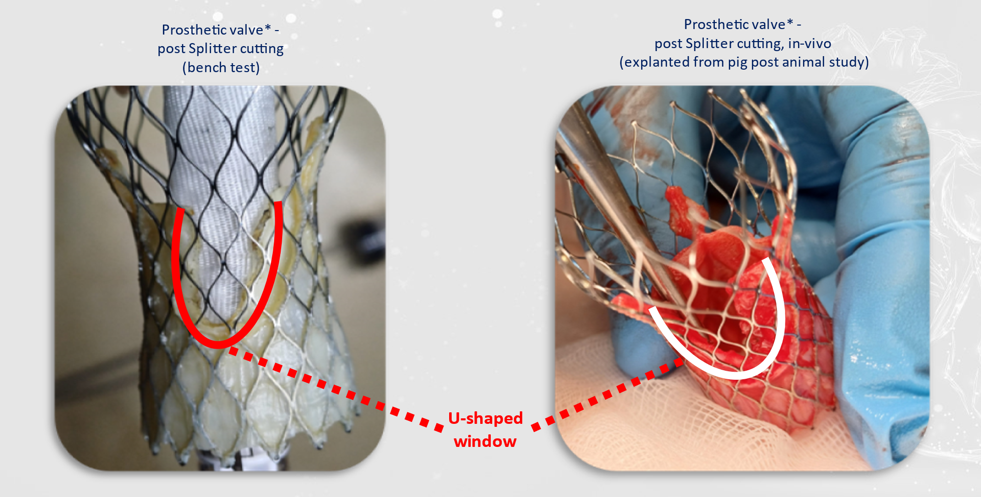
Note the large cut of the cusp, assuring adequate blood flow to the coronaries
The cut in the cusp of the existing valve, prior to TAV in TAV implantation
Benefits
Simple to operate
It enables precise control of the cutting site- no need in extreme pulling force to operate.
It allows the excision of a section (opening a window) of the heart valve and not just a straight cut.
Fully integrated into the workflow of the TAVI procedure.
Intuitive operation by interventional cardiology.
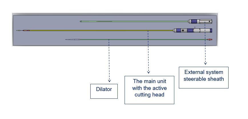
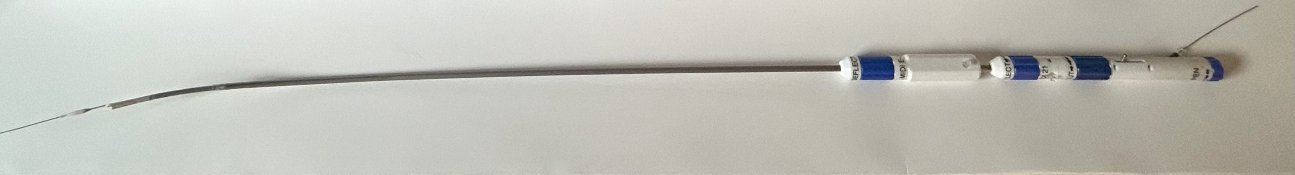
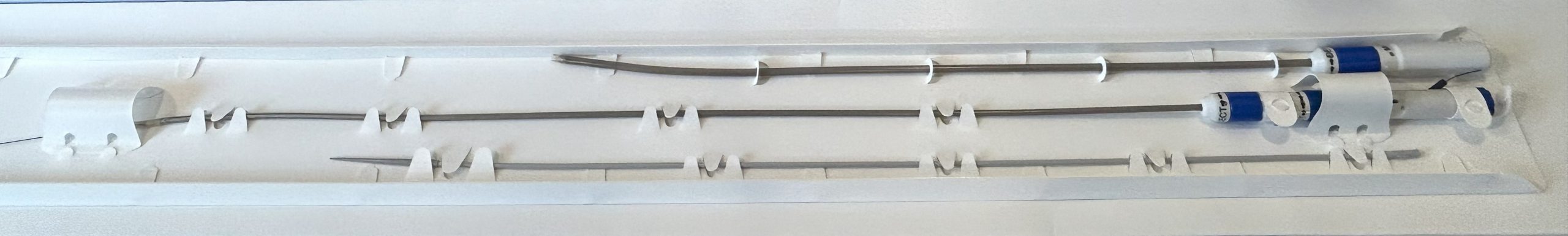
The two control units (on right) are designed to deliver and steer the cutting head across the aortic arch and grasp the selected leaflet of the aortic valve. Navigation is performed under Fluoroscopy and Echo imaging.
The 3 modules of the Splitter system are packed in a protecting tray. Each unit can be removed easily and assembled just prior to the procedure.
The unique advantages of the cusp resection by the cutting head are :
Excision of a large patch from the cusp, not just a slit
Removal of the patch out of the body
Providing positive confirmation to the doctor
Adequate blood supply to the coronaries
Enabling coronary access for future CSI procedures
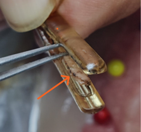
The jaws of the cutting head close firmly to hold the cusp, prior to the activation of the cutting wire. The excised tissue patch is secured by the jaws until the devices is pulled out of the artery
After excision, the doctor removes the Splitter device out of the artery. At the bedside, between the opened jaws of the cutting head, one can observe the excised tissue and thus receive positive confirmation, that the procedure was completed successfully.